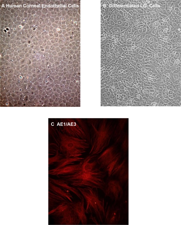
Figure 6.
Differentiation of rat LG immature cells in corneal endothelial cell media. Human corneal endothelial cells (A) and immature cells from LG (B) were grown in media developed for corneal endothelial cells for two weeks. Differentiated LG immature cells then were stained with the epithelial cell marker cytokeratin AE1/AE3 (C). Magnification 100× (A, B) and 200× (C).

