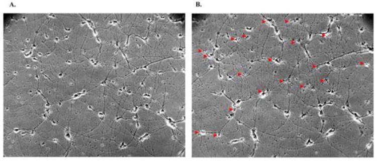
Figure 3.

Glutamate-induced neuronal death. Panel A illustrates the rat primary cortical neurons before glutamate treatment. Twenty-four hours after 10 minutes treatment with 100 μM glutamate, panel B shows that death occurred in many of the cortical neurons (arrows).
