Figure 6.
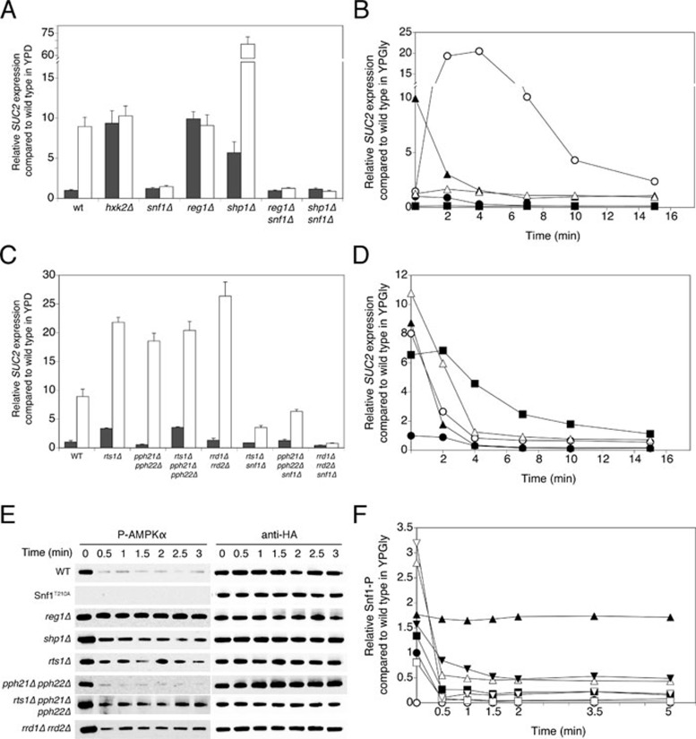
Requirement for PP1 and PP2A subunits in regulation of glucose repression. (A, C) Cells were grown in YPD medium containing 2% glucose (repressed cells, dark bars) and shifted to YPGly medium with 0.05% glucose and left to grow for another 3 h (derepressed cells, white bars). Samples were taken for determination of SUC2 expression level. (B, D) Cells were grown in YPGly medium with 0.01% glucose, at time point 0, 20 mM glucose was added. Samples were taken for determination of SUC2 expression level. (B) WT (•), reg1Δ (○), shp1Δ (▴), hxk2Δ (△) and snf1Δ (▪). (D) WT (•), rts1Δ (○), pph21Δ pph22Δ (▴), rts1Δ pph21Δ pph22Δ (△) and rrd1Δ rrd2Δ (▪). (E) Snf1 phosphorylation (Snf1-P) was detected by P-AMPKα antibody at the indicated time points after addition of 20 mM glucose to glucose-deprived (glycerol-grown) cells of specific deletion strains. Anti-HA loading controls are shown. (F) Snf1 dephosphorylation as shown in E was quantified and normalized (Snf1-P signal divided by anti-HA signal and normalized to the t = 0 time point of the WT strain). WT (•), Snf1T210A (○), reg1Δ (▴), shp1Δ (△), rts1Δ (▪), pph21Δ pph22Δ (□), rts1Δ pph21Δ pph22Δ (▾) and rrd1Δ rrd2Δ (▽).

