Figure 3.
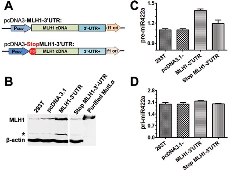
Expression of ectopic MLH1 protein increases the level of pre-miR-422a in 293T cells. (A) Plasmid pcDNA3.1 constructs carrying WT MLH1 cDNA and a mutant MLH1 gene whose ATG start codon was changed to a TAA stop codon by site-directed mutagenesis. (B) MLH1 expression in MLH1-deficient 293T cells transfected with pcDNA3.1 alone, pcDNA3.1 carrying WT or mutant MLH1, as indicated. Ectopic MLH1 protein was detected by western blotting, with β-actin serving as an internal loading control. (C, D) qPCR quantification of pre-miR-422a (C) and pri-miR-422a (D) in 293T cells transfected with the indicated plasmids. Data shown are from three independent experiments (mean ± SD). Statistical significance was determined by one-way ANOVA. *Degraded MLH1.

