Figure 3.
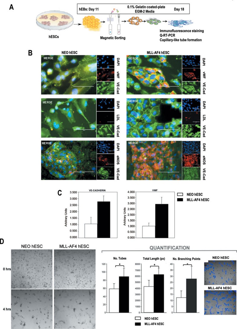
MLL-AF4 hemogenic precursors display an enhanced endothelial cell fate as compared to NEO hemogenic precursors. (A) Schematic of the endothelial differentiation and phenotypic and functional characterization of sorted hemogenic precursors. (B) NEO and MLL-AF4 hemogenic precursors isolated from day 11 EBs were cultured in EGM-2 media for 5-7 days and analyzed by immunohistochemistry for vWF, VE-cadherin and eNOS expression as well as LDL uptake (n = 3). Representative single stainings are shown on the right. A “merge” staining shows marker expression co-localization. (C) qPCR analysis of NEO and MLL-AF4 cells for the indicated endothelial-specific genes. This analysis was performed on NEO and MLL-AF4 hemogenic precursors after 6 days of culture in gelatin-coated plates, as indicated in A. (D) After 6 days of culture, NEO and MLL-AF4 hemogenic precursors were cultured in Matrigel at a density of 12 × 103 cells/well to assess their capacity to form capillary-like structures (n = 3). Images were captured 0 and 4 h after cell plating. Representative images are presented in the left panel. Quantification of the number of endothelial tubes, total length of tubes, and number of branching points was obtained with the software developed by Wimasis. Software-processed images are presented on the right as an example. Data are presented as mean ± SEM.

