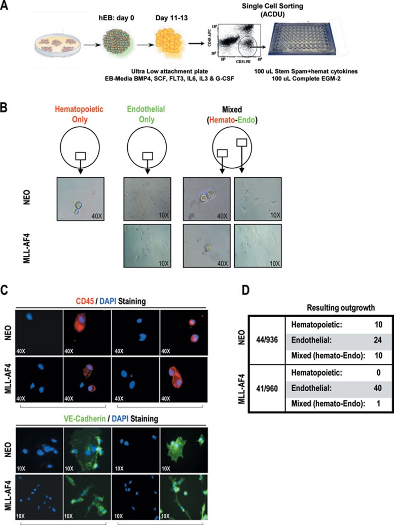
Figure 4.
Clonal analysis confirms an enhanced endothelial commitment of MLL-AF4 hemogenic precursors. (A) Schematic of EB development and FACS sorting of single hemogenic precursors into 96-well plates. (B) Phase contrast images showing hematopoietic outgrowth only, endothelial outgrowth only or mixed (hemato-endothelial) outgrowth. Hematopoietic cells are identified as small round refractive cells loosely attached to the gelatin-coated plastic whereas endothelial cells are larger spindle-shaped cells strongly attached to the gelatin-coated plastic. (C) The cells resulting from single hemogenic precursor proliferation in each individual well were analyzed in situ for CD45 (hematopoietic cell fate) and VE-cadherin (endothelial cell fate). CD45 expression is shown in red, VE-cadherin in green and DAPI nucleus staining in blue. (D) Summary table illustrating the resulting outgrowth within individual wells containing single NEO or MLL-AF4 hemogenic precursors. At 10 to 15 days after sorting, single wells were observed to contain (i) hematopoietic progeny only, (ii) endothelial progeny only or (iii) both hematopoietic and endothelial cells.

