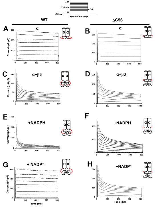
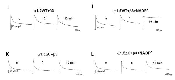
Fig. 2. Effect of Kv1.5 C-terminus deletion on Kvα-β currents.
Whole-cell currents recorded from COS-7 cells cotransfected with plasmids encoding WT or ΔC56 Kv1.5 alone or coexpressed with Kvβ3. Outward currents were recorded using the patch-clamp technique (n=6-9 cells each group). Panels show representative current traces recorded with pipette containing control internal solution from cells expressing Kv1.5WT (a); or Kv1.5ΔC56 (b); Kv1.5+β3 (c); Kv1.5ΔC56+β3 (d). Representative outward currents recorded from cells transfected with WT Kv1.5+β3 with patch pipettes containing 250 μ M NADPH (e) or 1mM NADP+(g) or cells expressing ΔC56 Kv1.5+β3 with 250 μM NADPH (f) or 1mM NADP+(h) in the patch pipette. The inset on the top panel shows the pulse protocol in which the membrane potential was held at −80 mV and outward currents were generated by depolarizing the cell from −60 mV to +60mV in 10mV increments for 800 ms. Inset to each trace is depicting the α with or without the β-subunit, The C-terminus of the Kvα-subunit extending from α to the β-subunit is shown in red. Representative traces shown in panels I-L are time course effects depicting the steady-state effects of NADP+. Panels I and K shows the currents from COS-7 cells co-expressing Kv1.5 or KvΔC with Kvβ3 patched with control internal solution. Panels J and L show cells patched with solution containing NADP+ (1mM).


