Fig. 3.
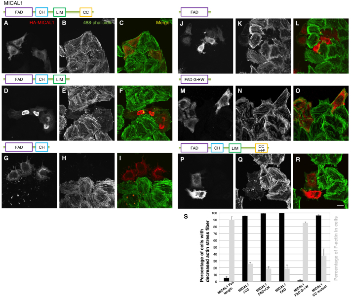
MICAL1 differs from MICAL2 and does not constitutively induce loss of actin stress fibers. (A–R) HeLa cells grown on coverslips were transiently transfected with either full-length HA–MICAL1 (A–C), truncated HA–MICAL1 (residues 1–799) lacking the CC domain (D–F), truncated HA–MICAL1 (residues 1–621) containing the FAD and CH domains (G–I), truncated HA–MICAL1 (residues 1–499) containing only the FAD domain (J–L), truncated HA–MICAL1 containing only the FAD domain but with residues 91–96 (GAGPCG) mutated to WAWPCW (M–O), or full-length HA–MICAL1 with residues 940–941 (AA) mutated to PP (P–R). After 18 hours, the cells were fixed, permeabilized and incubated with anti-HA antibody followed by Alexa-Fluor-568-conjugated anti-mouse secondary antibody and Alexa-Fluor-488-conjugated phalloidin. Scale bar: 10 μm. (S) Quantification of the effects described in A–R. A minimum of 100 transfected cells for each tranfection were used to calculate the percentage of cells displaying a decreased number of actin stress fibers (cells displaying fewer than five prominent stress fibers) compared with untransfected cells. F-actin levels were quantified from a minimum of 30 untransfected and transfected cells from each transfection of each MICAL construct. Quantification was performed using LSM5 Pascal software with data derived from three independent experiments. Error bars indicate s.e.m.

