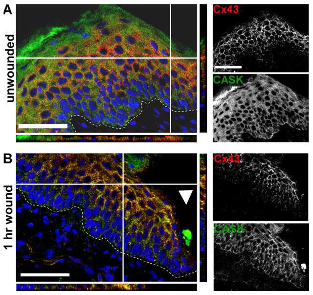
Fig. 4.

Human foreskin analyzed by immunostaining with antibodies to Cx43 and CASK. The x–y, x–z and y–z plane projections are shown in color and individual antibody staining is shown in grayscale. Cx43 staining is red and CASK staining is green. Nuclei were counterstained with DAPI (blue). Dashed line indicates basement membrane. Foreskins were wounded via punch biopsy and wound margin is indicated by white arrowhead. (A) Unwounded foreskin. (B) Skin at wound margin after 1 hour. Note that the CASK antibody, like many other antibodies, sticks nonspecifically to dead cells within the upper cornified layer, leading to the diffuse green staining. Scale bars: 50 μm.
