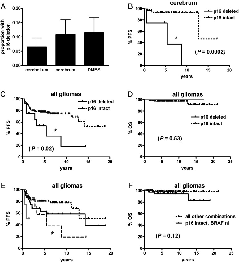
Fig. 4.
PFS and OS in pediatric low-grade gliomas by p16 and BRAF status. (A) There was no difference in the frequency of homozygous p16 deletion by tumor site (P = .64). While PFS was shorter in cerebral tumors (B) and all pooled gliomas (C) with p16 deletion, cerebellar and DMBS subsets did not achieve significance (see Results), nor did OS in all gliomas (D). (E) Stratifying all gliomas by p16 deletion and any BRAF abnormality (rearrangement or V600E mutation) showed that when BRAF was abnormal, the concomitant presence of p16 deletion correlated with shorter PFS (short dotted black line = p16 intact, BRAF abnormal; long dotted black line = p16 deleted, BRAF abnormal; solid grey line = p16 deleted, BRAF normal; solid black line = p16 intact, BRAF normal; *P = .04 vs p16 intact, BRAF abnormal). (F) There was, however, no significant stratification of OS by p16 and BRAF in this cohort. DMBS, diencephalon/midbrain/brainstem/spinal cord; nl, normal.

