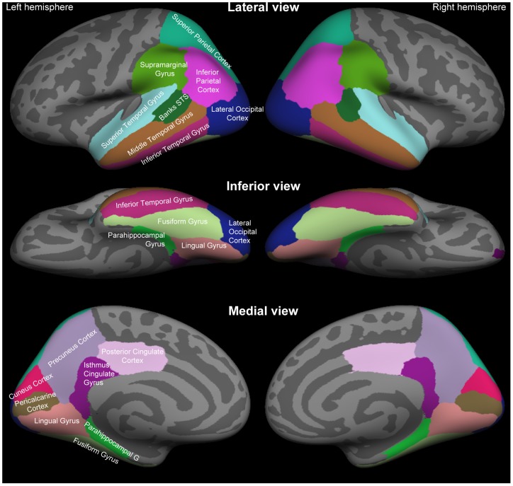
Figure 1. Standard anatomical parcellation of the posterior cortical surface.
Color-coded labels of anatomical ROI labels based on the Desikan-Killiany atlas [57] have been shown in the lateral (Top), inferior (Middle), and medial (Bottom) views of the FreeSurfer inflated standard-brain cortical surface. Abbreviation: STS, superior temporal sulcus.

