
Fig. 3.
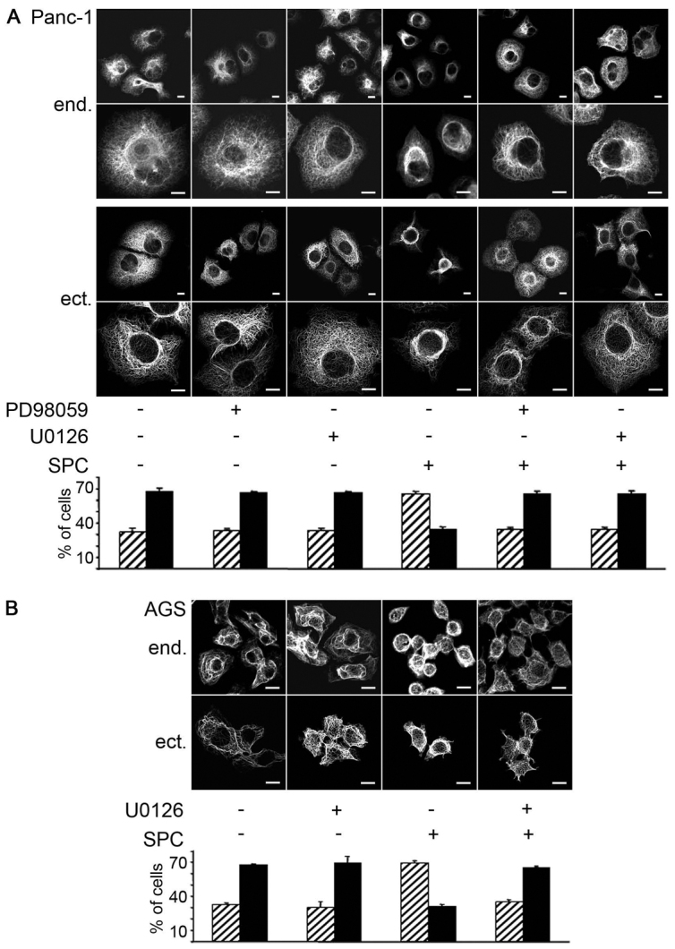
Role of p44 and p42 (MAPK1 and MAPK3) activation in SPC-induced keratin reorganization. (A,B) Cells were plated on coverslips and subsequently transfected with the respective plasmids for 48 hours. Panc-1 (A) and AGS (B) cells were incubated with either 15 μM PD98059 (only Panc-1) or 10 μM U0126 for 1 hour followed by 45 minute incubation with 15 μM SPC. Images of representative cells (two different magnifications in A) show endogenous (end.) stained with a pan-CK antibody, followed by Alexa Fluor 488 staining or transfected eCFP–K8(WT),eYFP–K18(WT) keratin (ectopic = ect.). Images were taken using a confocal microscope and keratin was detected within the 488 channel. Scale bars: 10 μm. In bar graphs in A and B, all cells on the coverslip or all transfected cells, respectively, were counted (between 50 and 200 cells per coverslip) and the ramified versus perinuclear phenotype of keratin was assessed by a person blinded for the specific condition. Data are expressed as the percentage of cells exhibiting a ramified or a perinuclear keratin phenotype and are the means ± s.e.m. of 3–10 independent experiments per condition.