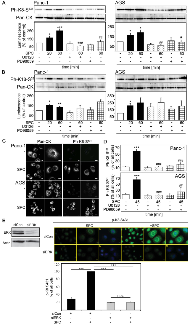
Fig. 4.
p44 and p42 (MAPK1 and MAPK3) mediate SPC-induced keratin phosphorylation. (A–B) Keratins were extracted from Panc-1 and AGS cells after treatment with 15 μM SPC and/or 15 μM PD98059 and 10 μM U0126. Keratin phosphorylation was analyzed by western blotting with antibodies against phosphorylated K8(S431) (Ph-K8-S431; A) or phosphorylated K18(S52) (Ph-K18-S52; B). Representative immunoblots are shown. Graphs display quantifications of luminescence. Error bars indicate the s.e.m. of at least four independent experiments. Significant differences were calculated using Student's t-test (* represents a significant difference between marked columns and untreated control; # represents a significant difference between marked columns and 60 minute SPC treatment). *,#P<0.05; **,##P<0.01; ***,###P<0.001. (C) Panc-1 and AGS cells were treated with 15 μM SPC for 45 minutes and keratin immunocytochemistry was performed using pan-CK and phosphorylated K8(S431) (Ph-K8-S431) antibodies, followed by Alexa Fluor 488 staining. Images show representative cells at two different magnifications. Scale bars: 10 μm. (D) Panc-1 and AGS cells were treated with 15 μM PD98059 or 10 μM U0126 for 1 hour followed by incubation with 15 μM SPC for 45 minutes. Immunocytochemistry was performed using a phosphospecific antibody against K8(S431) (Ph-K8-S431). Graph depicts quantification of pK8(S431)-positive cells compared with all stained cells (* represents significant difference between marked columns and untreated control; # represents significant difference between marked columns and 45 minute SPC treatment). **,##P<0.01; ***,###P<0.001. (E) p44 and p42 were depleted in Panc-1 cells using specific siRNA and confirmed by western blotting (left). p44/42 was depleted in Panc-1 cells followed by SPC treatment (20 minutes; 12.5 μM SPC) and keratin immunocytochemistry was performed using a phosphospecific antibody directed against phosphorylated K8(S431) (p-K8-S431) (right). Intense pK8(S431) immunoreactivity was detected exclusively upon incubation of cells with SPC, but not in unstimulated, control cells or cells in which p44 and p42 were depleted. pK8(S431) immunoreactivity was predominantly detectable in reorganized, perinuclear keratin filaments, indicating that K8 phosphorylation strictly correlates with keratin reorganization. Photographs show representative cells at two different magnifications. Scale bars: 10 μm. Graph depicts quantification of pK8(S431)-positive cells compared with all cells. Error bars indicate s.e.m. of three independent experiments. Significant difference was calculated using the Student's t-test. *P<0.05. M, merged image.

