Fig. 2.
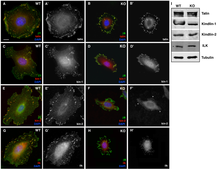
Localization of β1-cytoplasmic-domain-interacting proteins in WT and KO cells. In all panels, WT focal adhesions are stained with β1 (green), whereas KO focal adhesions are stained with β6 (green) and co-stained in red with talin (A,B), kindlin-1 (C,D), kindlin-2 (E,F) and ILK (G,H). Talin and kindlin-1 strongly localize to the focal adhesions of both cell types (A′–D′). Kindlin-2 fails to localize to the focal adhesions in the KO cell (arrowheads in F′) and there is very poor recruitment of ILK to the focal adhesion in the KO cells (H′). (I) Western blot analysis of WT and KO lysates with talin, kindlin-1, kindlin-2 and ILK. Tubulin was detected as a loading control. Scale bar: 10 μm.

