Fig. 8.
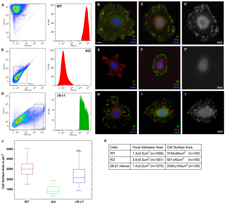
Recruitment of Kindlin-2 by β6 integrin mediates cell spreading and focal complex formation. (A) Surface expression of endogenous β1 integrin on WT cells assessed by FACS. (B,C) WT cells stained for β1, vinculin and kindlin-2 (C′) as indicated. (D) Surface expression of endogenous β1 integrin on KO cells assessed by FACS. (E,F) KO cells stained with β1, vinculin, β6 and kindlin-2 (F′) as indicated. (G) Cells expressing high levels of β6-β1 were sorted based on high expression of GFP. Cells rescued with β6-β1 (H,I) were stained with β6 vinculin and kindlin-2 (I′) as indicated. (J) Box plot of the distribution of the average cell area along the median (n=100) of WT, KO and β6-β1-rescued cells (***P<0.001, compared with KO cells). (K) Table showing the mean focal adhesions and cell surface areas of the WT, KO and β1-β6-rescued cells. Scale bar: 10 μm.

