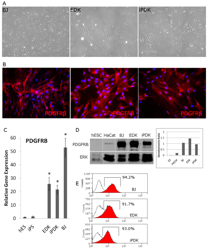
Fig. 1.
Expression of PDGFRβ in ESC- and iPSC-derived cells correlates with mesenchymal phenotype. Cells differentiated from ESCs (EDKs) and iPSCs (iPDKs) were morphologically similar to control fibroblasts (A) and expressed PDGFRβ as detected by immunofluorescence staining (B). (C) Analysis of mRNA levels of PDGFRB by real-time RT-PCR, showed that EDK, iPDK and BJ fibroblasts expressed significantly higher levels of PDGFRB compared with pluripotent cell types (ESCs and iPSCs). Error bars represent s.d. *P≤0.05 compared with ESCs. (D) Western blot analysis showed that PDGFRβ is similarly expressed in BJ, EDK and iPDK cells, is not expressed in ESCs and is expressed at very low levels in an unrelated epithelial cell line, HaCat. (E) These results were also confirmed by flow cytometry showing that greater than 90% of EDK and iPDK cells expressed detectable levels of PDGFRβ at the cell surface, similar to control fibroblasts (BJ).

