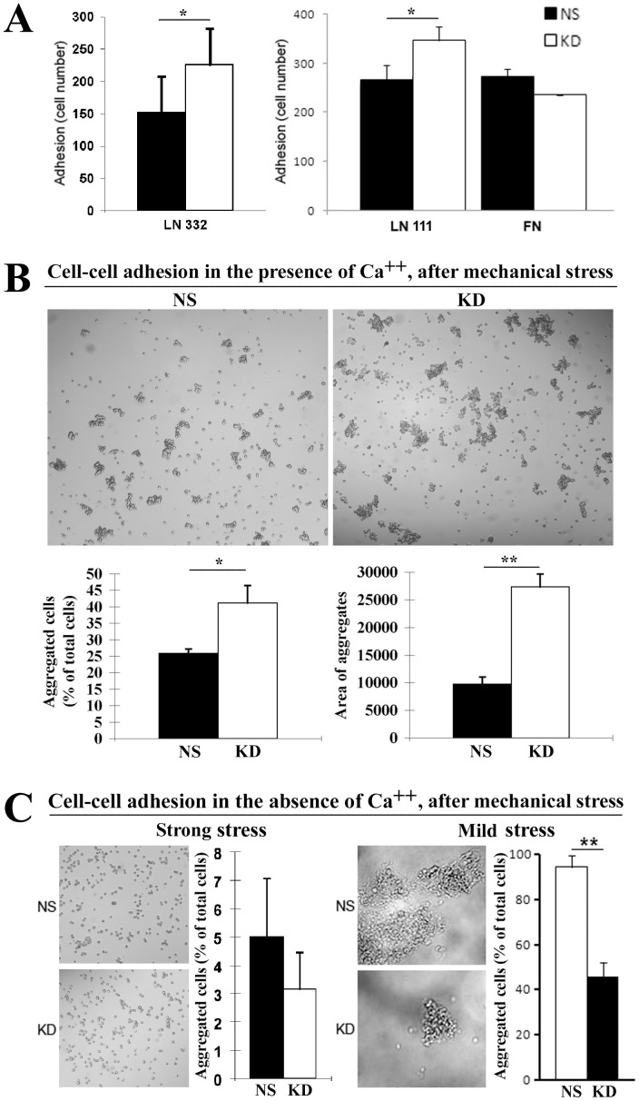
Figure 3. CO-029 regulated cell-matrix and cell-cell adhesions.
(A) Cell-matrix adhesion. Adhesion onto laminin 111, laminin 332, and fibronectin of HT29-NS and -KD cells was assayed after incubating at 37oC in 5% CO2 for 1 h. The adhesion of the KD cells on laminin 111 and laminin 332 was significantly increased compared with NS cells (P = .042 on laminin 111 and = .048 on laminin 332). (B) Total cell-cell adhesion. Cell aggregation was measured in the Ca++-containing media after a relatively strong mechanical force was applied. (C) Ca++-independent cell-cell adhesion. Cell aggregation was measured in the Ca++-free media after relatively strong or mild mechanical forces were applied, respectively. The aggregation of NS and KD cells after shear stress were quantified as described in Materials and Methods. P values are 0.03 for the aggregated cells in the presence of Ca++, 0.001 for the area of aggregates in the presence of Ca++, and 0.0004 for mild stress in the absence of Ca++. Images of cell aggregates after the shear-stress treatment were obtained under phase-contrast microscopy. All of the data are projected as mean±SEM (n = 4). *P<.05, **P<.01.

