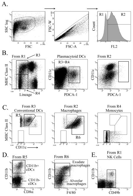
Figure 1. Identification of pulmonary mononuclear phagocytes by flow cytometry.
(A) Gating strategy for lung cell suspensions based on size, doublet discrimination and low (R1) and high (R2) autofluorescence, respectively. (B) Representative dot plots illustrating gating strategy to define pDCs. Note pDCs are absent from the high autofluorescence gated population (right). (C) Representative dot plots illustrating gating to define cDCs, macrophages and monocytes. (D) Dot plots illustrating further breakdown of cDCs into CD11b+ and CD11b− subsets and macrophages into exudate and alveolar macrophages. (E) NK cells gated from R1 were identified as CD3− CD49b+ cells with or without CD11b expression.

