
Figure 1.
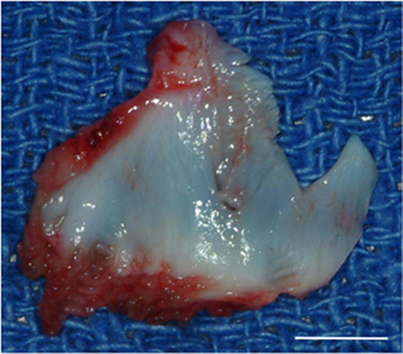
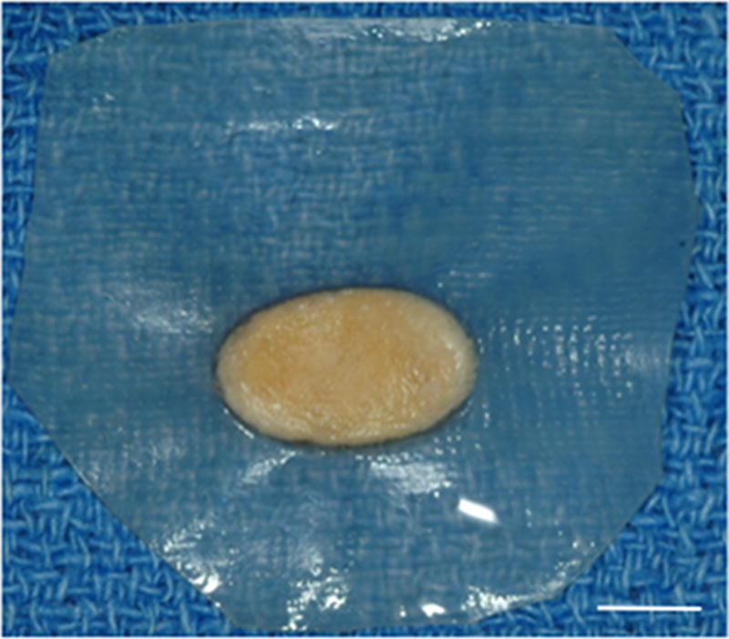
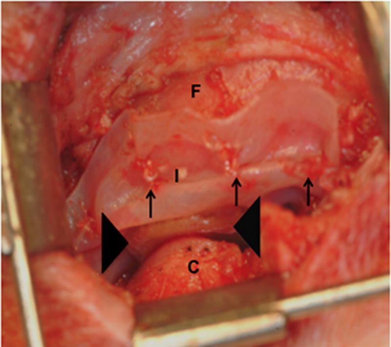
Images showing the surgical procedure. The native disk (A) is exposed and excised leaving the joint space empty. UBM implants (B) were hydrated and trimmed to size prior to placement between the condyle and fossa on the experimental side and attachment through the fossa (C). Note sutures through the anchoring site (arrows) and interpositional pillow-like core (arrowheads). The contralateral side was left devoid of a meniscal substitute. C = condyle, F = fossa, M = disk, and I = implanted UBM-ECM device. Scale bars in A and B = 5 mm.