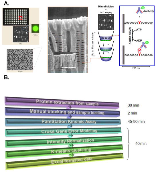
Figure 1. PamGene Array Technology.
A) The PamChip®96 Array contains 96 spots/chip with each spot on the microarray containing up to 400 putative substrates allowing for multiplex analysis. The array is made of a porous aluminum oxide material 60 μm thick with interconnected capillaries having 500 X surface area of 2D arrays. This shows an enlarged schematic of the capillaries with peptide substrates indicated. In this example, the substrates are peptide sequences containing consensus tyrosine phosphorylation sites representing the tyrosine kinome. Lysates containing active kinases are pumped through the pores and phosphorylate specific peptides. The degree of phosphorylation is measured in real time by fluorescently labeled phospho-antibody binding detected by CCD cameras. See Materials and Methods for additional detail. B) PamStation® flow chart with estimated time required: Sample preparation requires only 30 minutes of time. The actual microarray analysis utilizes manual blocking of the chip and application of the sample (requiring about 2 minutes) followed by a fully automated analysis by the PamStation® (requiring 45–90 minutes). Lastly, data analysis is also automated such that annotated and quantified raw data, initial velocities and end levels, and coefficient of correlation for each readout can be obtained in exportable format (such as Excel) within 40 minutes.

