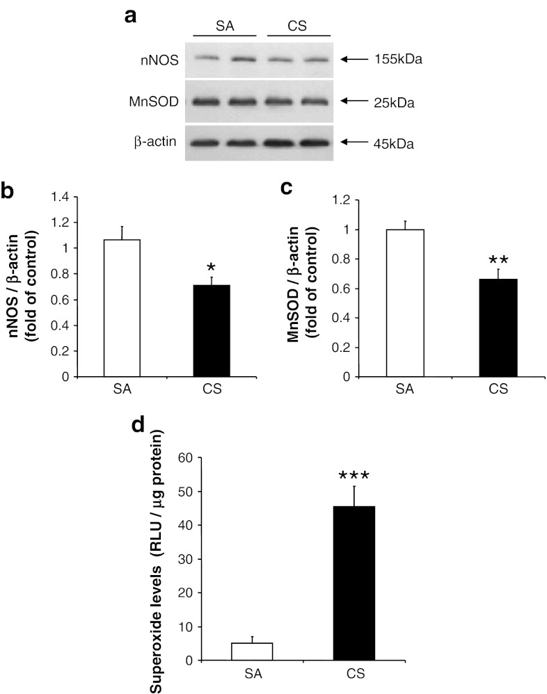
Fig. 4.
Changes in nNOS, MnSOD protein levels, and superoxide levels after 56 days of CS exposure. a Protein levels of nNOS and MnSOD, with β-actin serving as an internal control. b, c Quantitative analysis of band intensity from the blots. Results are expressed as fold of control. d Superoxide level measured in cerebral cortex. Values represent the mean ± SEM. *p < 0.05, **p < 0.01, *** p < 0.001, when comparing between SA and 56 days of CS exposure groups (n ≥ 7)

