Figure 6.

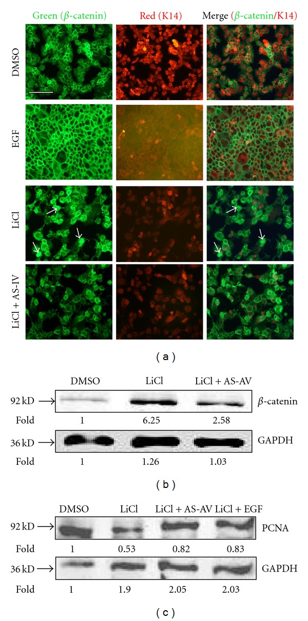
β-catenin is differentially regulated by LiCl and AS-IV. Stabilization of β-catenin in keratinocytes was followed by immunofluorescence staining with a β-catenin-specific antibody (a) and Western blot analysis (b). (a) DMSO (negative control) and EGF (positive control) treated keratinocytes exhibited normal β-catenin membrane expression. The LiCl-treated group showed stabilized β-catenin, which was downregulated following AS-IV treatment. (b) GAPDH protein expression was used as the protein loading control. (c) Western blot analysis revealed that PCNA expression, which is related to cell proliferation, was downregulated 50% by LiCl compared to the control cells, and upregulated to 80% by AS-IV. GAPDH expression was used as a protein loading control. Asterisks indicate statistically significant differences between control and LiCl-treated cells. The scale bar in the first panel of (a) represents 100 μm, and is applicable to both sections.
