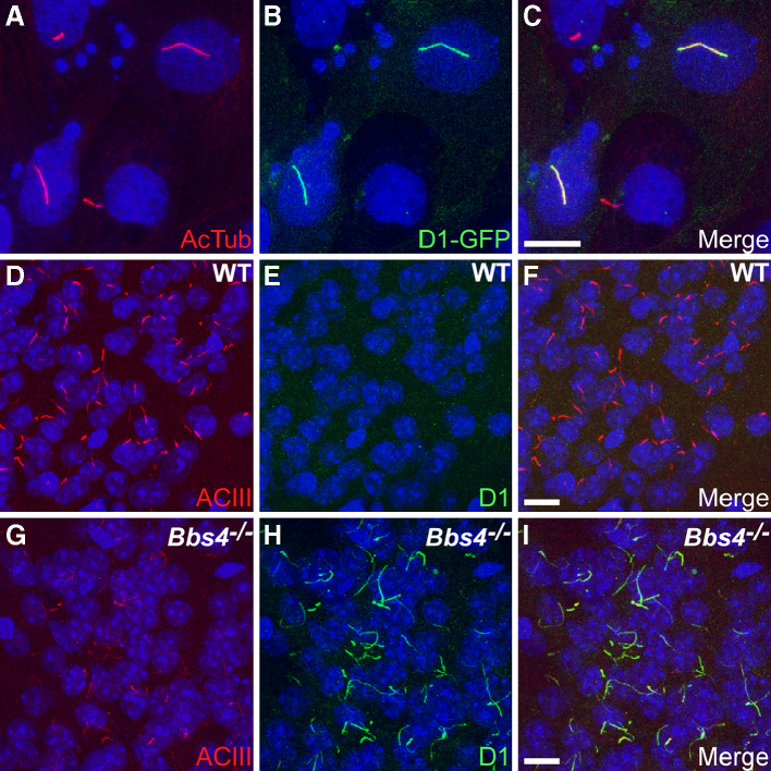
Fig. 1a–i.
Dopamine receptor 1 (D1) localizes to cilia, and ciliary localization is increased in the brains of Bbs4 −/− mice. a–c Representative image of transiently transfected inner medullary collecting duct (IMCD) cells expressing D1 fused at the C-terminus to EGFP. a Acetylated α-tubulin (AcTub; red) marks the cilia; b EGFP fluorescence (green) shows expression of the D1 receptor; c merged images. Representative images of the basolateral amygdala in adult WT (d–f) and Bbs4 −/− (g–i) mice (n = 3 animals for each genotype) showing labeling for type III adenylyl cyclase (ACIII; red) and D1 (green). Nuclei are stained with DRAQ5 (blue). The appearance and distribution of ACIII-positive cilia is similar between WT (d) and Bbs4 −/− (g) sections. The identical fields showing labeling for D1 (green) reveal a lack of D1-positive cilia in the WT (e) section but abundant D1-positive cilia in the Bbs4 −/− (h) section. Merged images showing no D1 labeling of cilia in the WT (f) section and colocalization of ACIII and D1 to cilia in the Bbs4 −/− (i) section. Scale bars represent 10 μm

