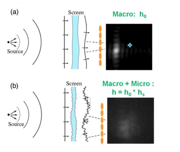
Fig. 2.

Schematic diagram of an aberrated eye viewing (a) without a random screen, (b) through a random screen and sample spot images. Ocular sources of scatter are modeled by a thin random screen in the plane of the eye’s pupil. The lenslet arrays are colored in orange. Courtesy of John. R. Hoffman (Lockheed Martin) during the workshop at Institute for Mathematics and Its Applications (IMA) at University of Minnesota.
