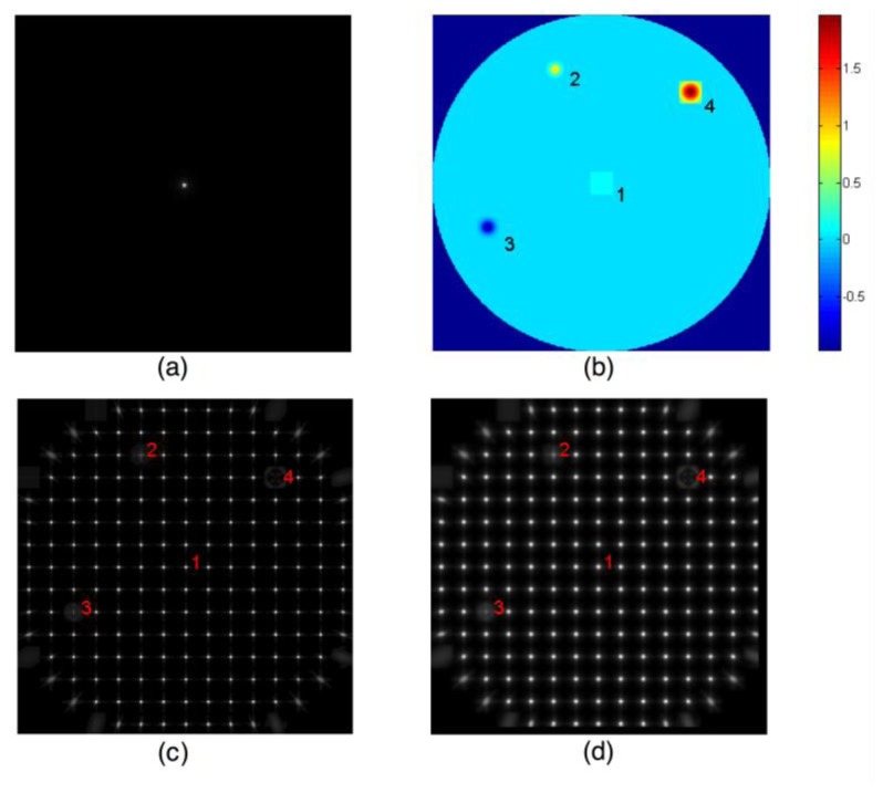
Fig. 5.

Graphical representation of scatter analysis in the double pass setup for Gaussian xerop. (a) PSF on the retina from the first pass is the object to be imaged on the second pass. Pixel size = 2.72 arcmin. (b) Beam location (1) and several Gaussian xerops (2-4). The Gaussian perturbation at location 3 is a drop of wetness that increases optical path length. The Gaussian xerops at locations 2 and 4 represent thinning of the tear film that shortens the optical path length. (c) PSFs for the second pass. Pixel size = 0.97 arcmin. (d) Double pass SH image. Pixel size = 0.97 arcmin. Note that the PSFs in (c) are computed for a point source on the retina. Since the retinal image formed from the first pass will contain blur to become an extended object for the second pass, the SH images in (d) are not strictly PSFs. For display, a square-root transformation was applied to the computed image.
