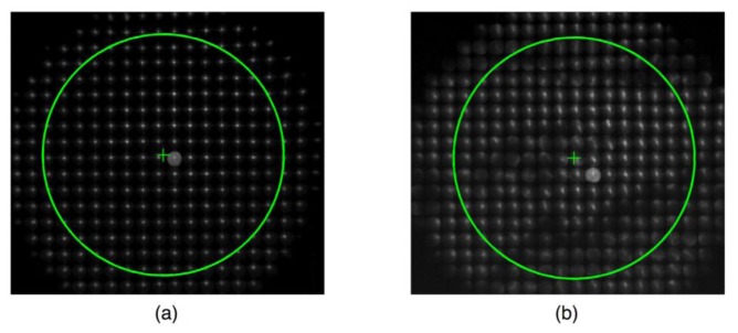
Fig. 7.
The raw image of the SH spots encircled with a pupil of radius 3 mm. The diameter of each lenslet is 400 microns in the SH detector. We expect a 15x15 array of the spot images inside the pupil. The ' + ' sign indicates the pupil center, which does not necessarily coincide with a spot in a lenslet. The intensity of the raw images was boosted for the display purpose. (a) the baseline data when the tear film forms a smooth surface (soon after ablink), (b) the SH image after the tear break-up (following blink-suppression).

