
Figure 6.
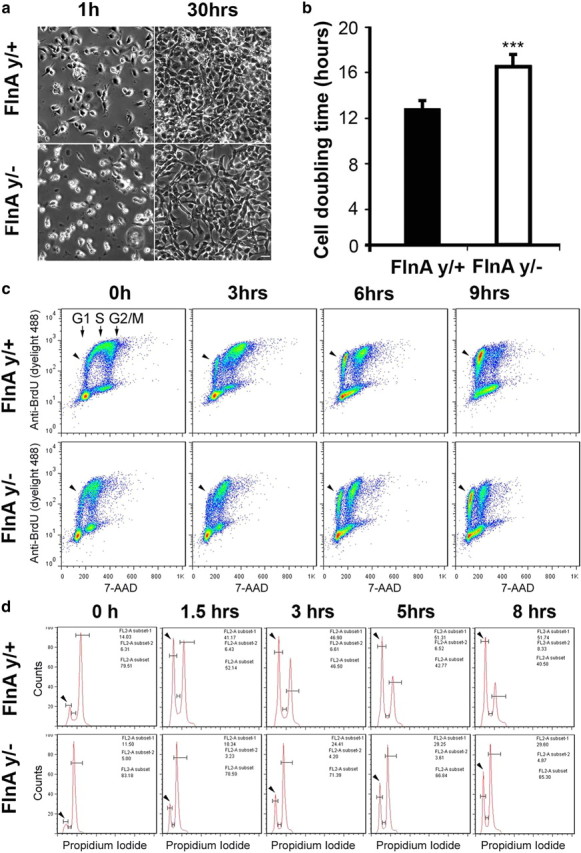
FlnA regulates cell cycle progression from G2 to M phase. a, FlnAy/− progenitors underwent a slower rate of growth as visualized with phase contrast microscopy. Neural progenitors from E13.5 brain cortex were plated on laminin-coated dishes at a cell density of 5 × 10⁴ cells/cm2. FlnAy/+ progenitor cells grew to confluency within 30 h after plating, whereas FlnAy/− progenitor cells achieved only 70–80% confluency during the same time interval and under the same culture conditions. Scale bar, 50 μm. b, Quantification to the right indicates a 25% decrease in the proliferation rate for FlnAy/− neural progenitors compared to progenitors isolated from littermate controls (n ≥ 3 independent samples per variable; ***p < 0.001). c, Flow cytometric analyses demonstrated a faster cell cycle progression from S to G1 phase in FlnAy/+progenitors. The cell cycle of BrdU-labeled progenitors progresses from S to G1 phase (arrowheads) for varied culture time post BrdU pulse. Cultured progenitor cells were pulse labeled with BrdU for 40 min and fixed at various time points after pulse. The fixed cells were stained with anti-BrdU antibody and 7-AAD, a fluorescent dye for DNA. d, Flow cytometric analyses showed that FlnAy/+progenitors underwent a faster cell cycle progression from G2/M (second unlabeled peak to the right) to G1 (arrowheads) phase. Cultured neural progenitors were synchronized to G2/M phase by nocodazole treatment and then released from nocodazole to allow cycle progression. The cells were harvested at various time points and stained with PI. Sample flow cytometry results were shown for n > 3 replicates (p < 0.001).