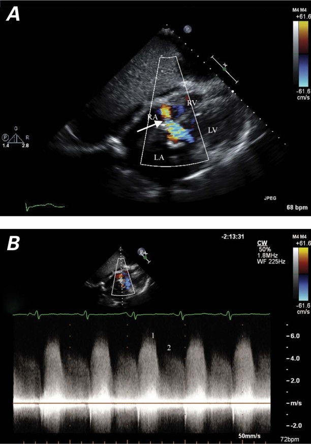
Fig. 4 A) Color-flow Doppler echocardiogram (subcostal view) shows an aortic-insufficiency jet entering the right atrium through the Gerbode defect (arrow) during diastole. B) Continuous-wave Doppler echocardiogram through the Gerbode defect shows a higher velocity of flow entering the right atrium during systole (1), and a lower velocity of aortic-insufficiency flow entering during diastole (2).
LA = left atrium; LV = left ventricle; RA = right atrium; RV = right ventricle
