Figure 4.
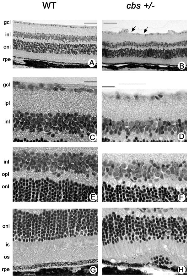
Histology of retinas of 30 week old heterozygous and wildtype mice. Low magnification light micrographs of hematoxylin and eosin-stained retinas from (A) WT (cbs+/+) and (B) heterozygous (cbs+/−) mice. Higher magnification light photomicrograph images of inner (C,D), middle (E,F) and outer (G,H) retina of WT and cbs+/− mice, respectively. Generally, the laminar organization of the retina in cbs+/− mice is similar to that of WT mice, although the inner plexiform layer is slightly reduced in thickness and there is clear evidence of dropout of ganglion cells (arrows in panel B) in these retinas. There are occasional aberrations observed in the region of the RPE as shown in panel H. Calibration bar = A, B = 50 μm, C-H = 20μm. Abbreviations: gcl = ganglion cell layer, ipl = inner plexiform layer, inl = inner nuclear layer, opl = outer plexiform layer, onl = outer nuclear layer, is = inner segment, os = outer segment, rpe = retinal pigment epithelium.

