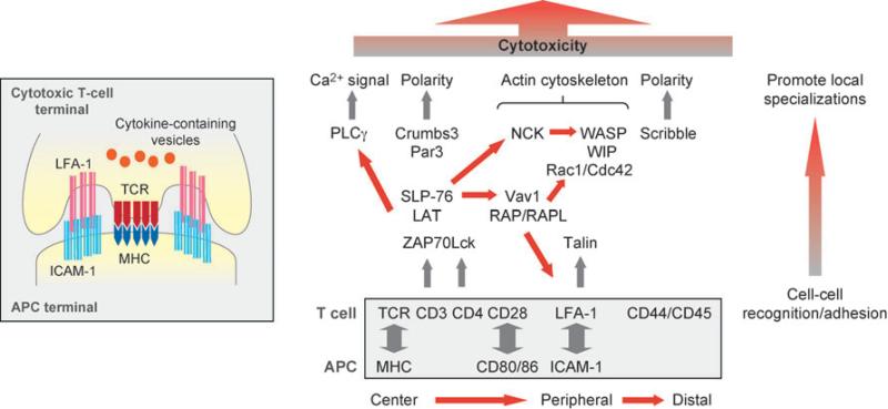
Figure 4.
Schematic representations of the immunologic synapse between T cells and antigen-presenting cells (APCs) (inset), adhesion proteins (boxed), and protein networks assembled at the synapse that remodel the T cell and initiate the immune response from the T cell. Abbreviations: ICAM-1, intercellular adhesion molecule 1; LAT, linker for activation of T cells; LFA-1, lymphocyte function-associated antigen 1; MHC, major histocompatibility complex; Par3, partition defective 3; PLCγ, phospholipase C γ; SLP-76, Src homology 2 domain-containing leukocyte-specific phosphoprotein of 76 kDa; TCR, T-cell receptor; WASP, Wiskott-Aldrich syndrome protein; WIP, WASP-interacting protein; ZAP70, zeta-chain-associated protein 70.

