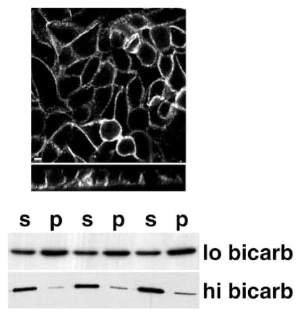
Fig. 3.
Effect of sodium bicarbonate concentration on Sec6/8 complex distribution. Confluent MDCK cultures on polycarbonate filters were grown in DMEM containing either 1 g/l (‘lo bicarb’) or 3.7 g/l (‘hi bicarb’) sodium bicarbonate for 48 hours. (Top) Cultures were fixed with 4% paraformaldehyde before extraction with buffer containing 1% Triton X-100. Anti-Sec6 monoclonal antibody (9H5) was visualized with FITC-labeled goat anti-mouse antibody. Confocal images were obtained as described in Fig. 1 legend. Scale bar: 5 μm. (Bottom) Triplicate filters of cells grown in hi or lo bicarbonate were extracted successively in Triton X-100 and SDS, as described in Materials and Methods. Sec8 in Triton-soluble (‘s’) and Triton-insoluble (‘p’) fractions was quantified by SDS-PAGE and western blotting. Protein levels were quantified using a Molecular Dynamics Phosphorimager. In 1 g/l bicarbonate, Sec8 is enriched at the apical junction (Fig. 1) and is only partially (~30%) soluble in Triton X-100. In 3.7 g/l bicarbonate, Sec8 is diffusely distributed along the lateral and basal membranes and is almost entirely (~90%) soluble in Triton X-100.

