Figure 5.
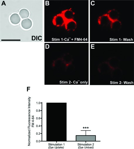
Isolated bead−presynaptic complexes are capable of recycling synaptic vesicles. The membrane dye FM 4-64 was used to visualize synaptic vesicle recycling. (A−E) Representative image panel of a cluster of three bead−presynaptic complexes (A) that have been stimulated using HBSS containing FM4-64 dye and 10 mM Ca2+ (Stim 1; B,C). This stimulation triggers SV fusion with the plasma membrane and internalization of the dye via incorporation into SV membranes. The second stimulation involves the use of HBSS with Ca2+ alone (Stim 2; D,E). In panels B and D, the images were acquired during stimulation, while in panels C and E, the images were captured during the wash (rest) phase of the protocol. (F) Quantification of the fluorescence changes in response to uptake (Stim 1) and unload (Stim 2) of the dye. The ∗∗∗ indicate p < 0.0001, n = 14 beads pooled from at least four independent coverslips. Scale bar, 10 μm.

