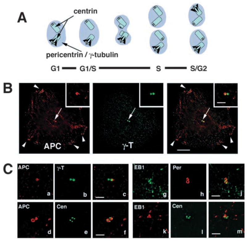
Fig. 1.

Preferential localization of APC and EB1 to a subset of centrioles. (A) Schematic representation of centrosome duplication during the cell cycle and localization of centrosome marker proteins. Pericentrin and γ-tubulin localize to the pericentriolar material (lilac area); centrin localizes to the centrioles (light blue tubes). MT-anchoring appendages on the mother centriole are marked in black. (B) Basal section of a MDCK cell co-stained for APC (red) and γ-tubulin (green). Arrowheads mark cortical APC clusters, arrow marks localization of APC with γ-tubulin. Bar, 10 μm. Insets show another example of the centrosome region of an MDCK cell at higher magnification. Bar, 2 μm. (C) Sections of U-2 OS cells showing the centrosome regions of cells immunostained for γ-tubulin (green in a–c), pericentrin (red in g–j) and centrin (green in d–f,k–m) and co-stained for APC (red in a–f) and EB1 (green in g–m). Cells with two centrosomes marked by pericentriolar pericentrin (red in g–j) or γ-tubulin (green in a–c) or with four centrioles marked by centrin (green in d–f,k–m) are also shown. APC and EB1 localize preferentially to one of two centrosomes (a–c for APC, g–j for EB1) and to two of four centrioles (d–f for APC) or to one of four centrioles (k–m for EB1). See also Movie 1 (http://jcs.biologists.org/supplemental). Bar, 2 μm.
