Fig. 8.
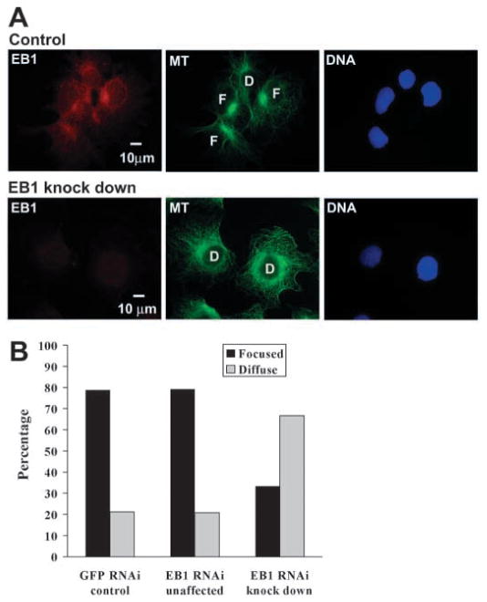
Depletion of EB1 with small interfering RNA reduces MT minus-end anchoring at the centrosome. (A) Cos-7 cells incubated with siRNA against GFP as a control or with siRNA against EB1 were immunostained for EB1 (red), and α-tubulin (green) and co-stained with DAPI for DNA (blue). Immunofluorescence images of EB1 were taken with identical exposure times to allow comparison of fluorescence intensity between images. Examples of cells with MT minus ends focused at the centrosome are marked as ‘F’ and examples of cells with reduced MT minus-end focus are marked as ‘D’ for diffuse. (B) The number of ‘F’ and ‘D’ type cells with EB1 knock down was quantified and compared with the number of these cell types in control cultures (‘GFP RNAi control’). Knock down was defined by reduced immunostain for EB1 (‘EB1 RNAi knock down’) and cells in the EB1 siRNA-treated cultures that did not show EB1 depletion were quantified as an internal control (‘EB1 RNAi unaffected’). Cells depleted for EB1 show reduced focus of MT minus ends at the centrosome. GFP RNAi control, n=165; EB1 RNAi unaffected, n=101; EB1 RNAi knock down, n=108.

