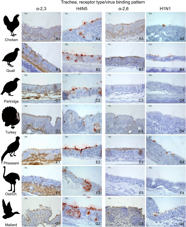
Figure 2.
Influenza receptor distribution and pattern of viral attachment in the trachea. Composite bright field microscope images comparing the distribution of α-2,3 and α-2,6 receptors, demonstrated by means of MAAII and SNA lectin histochemistry, with the pattern of viral attachment of the avian influenza A/Mallard/Netherlands/13/08 (H4N5) virus and the human influenza A/Netherlands/35/05 (H1N1) virus, demonstrated by means of virus histochemistry, in the trachea of chicken (A1-A4), common quail (B1-B4), red-legged partridge (C1-C4), turkey (D1-D4), golden pheasant (E1-E4), ostrich (F1-F4), and mallard (G1-G4).

