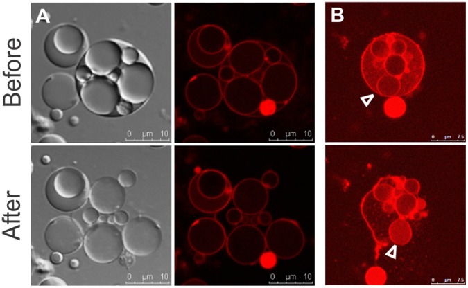
Figure 4. Release of internal vesicles.
(A) Following collapse of an L-form mother cell, intact progeny cells are released. Membranes are visible by using DIC (Differential Interference Contrast) microscopy (left); dye-labeled membranes (right) are shown immediately before (upper) and after the collapse (lower). (B) A vesiculated cell labeled with Rho123 (upper) disintegrates and releases a vesicle (lower image, arrow head). The increase in the Rho123 signal on the surface of the released vesicle suggests an increase in membrane charge.

