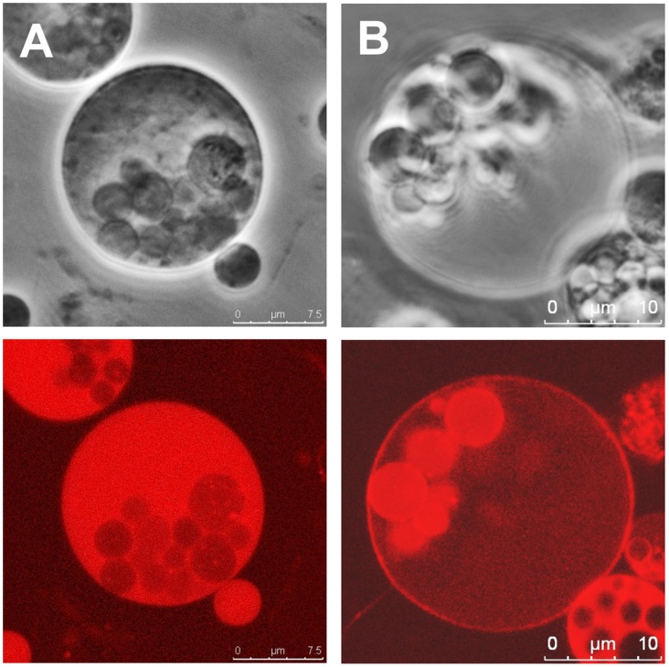
Figure 7. Enterococcus L-form cells.
E. faecium (A) and E. faecalis (B) L-form cells were generated and stained with Rho123 (10 µg ml−1) to indicate charged membranes. In contrast to Listeria L-forms, the intracellular vesicles of Enterococcus L-forms still enclosed in the mother cell cytoplasm frequently feature stronger phase contrast (upper panels) and more intense Rho123 accumulation and fluorescence (lower panels).

