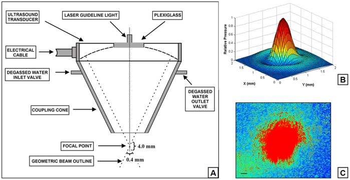
Figure 2. Schematic diagram of the transducer assembly. A.
: Spherical transducer with coupling cone and a centrally attached laser guideline light. The ellipsoidal acoustic focal zone covers approximately 4 mm axially and 0.4 mm transversely at the half-pressure points. B: Normalized pressure distribution through the culture dish for the single element on the radial plane at the distance of 62.75 mm of the transducer’s surface. C: The focal point of focused ultrasound guided by red laser light in the middle field of view of the microscope objective. Scale bar, 50 µm.

