
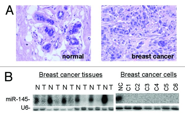
Figure 1. Downregulation of miR-145 in human breast cancer. (A) Pathological examination of normal breast tissues and breast tumor tissues using H/E staining. Normal breast tissue consisted with well-differentiated gland cells and milk ducts. Magnification: 200× . (B) Northern blot analysis shows that miR-145 is expressed at high levels in normal breast tissues (N) but at very low levels in breast cancer tissues (T, left panel) and breast cancer cell lines (right panel). C1 to C6 represent MCF7, ZR-75–30, T47D, MDA-453, MDA-435 and MDA-231 cells.