
Figure 1. Structural analysis of Hla.
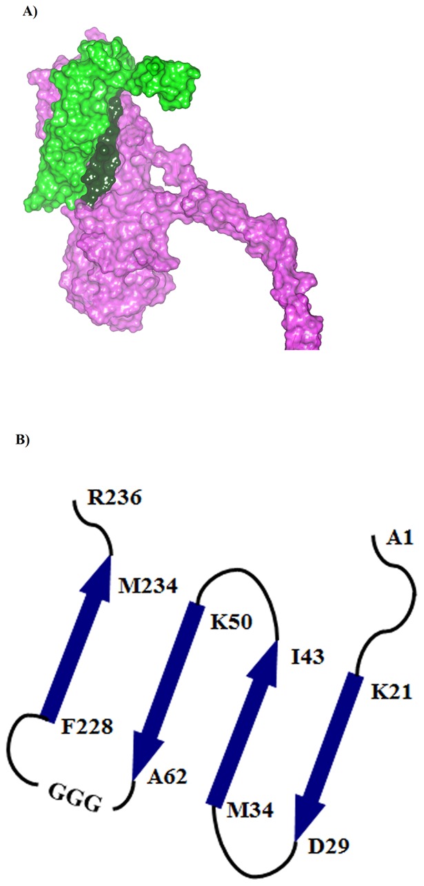
(A) The relative topology of 1–62 and 1–62(GGG)–(223–236) AT constructs on the protein surface of a subunit from the 7AHL heptameric hemolysin crystal structure. The protein surface for the 1–62 segment is colored green, the 223–236 sequence colored dark green, and the remaining structure colored purple. (B) Topology of the secondary structural elements in α-hemolysin for peptide segments examined in this study.