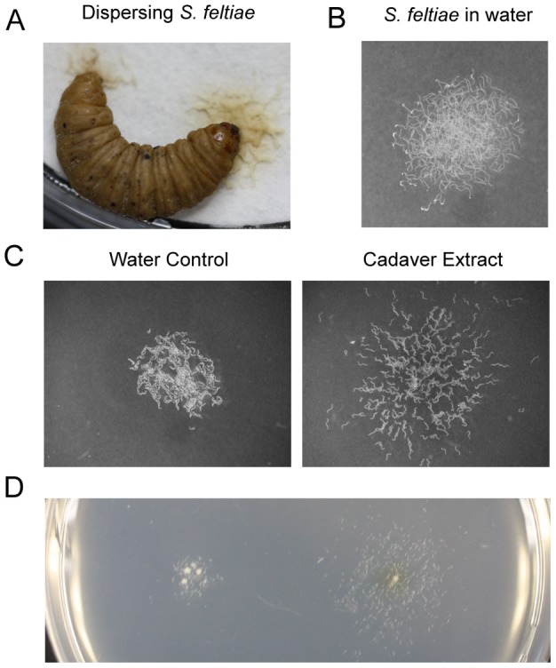
Figure 1. Dispersal assay.

(A) Naturally dispersing infective juveniles (IJ) of S. feltiae from an insect cadaver. (B) Approximately 300 IJs were placed on an agar plate in water. (C) IJs were treated with either water (control) or insect cadaver extract. Images are representative of six experiments for each treatment. (D) Dispersal assay on the same plate. The illustration represents two experiments. Behavior is temperature and season dependent. Assays were conducted at RT (22±0.5°C) or under temperature controlled conditions.
