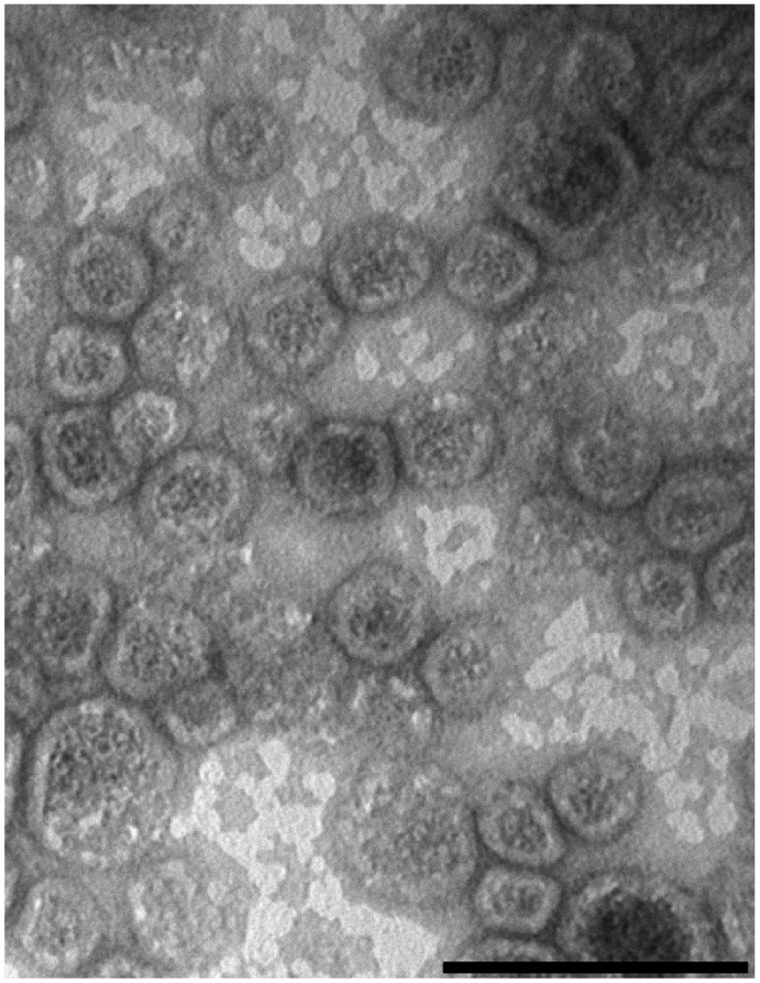
Figure 3. Electron microscopy of Shigella sonnei ΔtolR ΔgalU GMMA.
GMMA were isolated from the culture supernatant of S. sonnei –pSS ΔtolR ΔmsbB by TFF, prepared for negative staining, and viewed by electron microscopy revealing the presence of well-organized membrane vesicles with a diameter of about 30–60 nm. Bar length = 100 nm.

