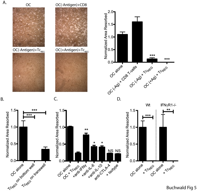
Figure 5. TcREG inhibit osteoclast activity in an antigen- and contact-independent manner by secreted cytokines.
A. Osteoclasts were seeded on hydroxyapatite-coated plates and allowed to adhere overnight. The osteoclasts were pulsed with SIINFEKL peptide (+Antigen) or a control FLAG peptide (-Antigen). OT-I TcREG or naïve CD8 T-cells were added and co-cultured for 7 days. Cells were re-fed with medium containing M-CSF and RANKL every 2 to 3 days. The plates were then treated with bleach solution, washed, dried and photographed. Representative photomicrographs are shown on the left. Quantitation from four experiments of three wells each is shown on the left. B. TcREG were added to top-insert of transwell (0.45-µ membrane) separated from the osteoclast plated on 24-well Corning Osteo-Assay plate. Osteoclast activity was determined by quantifying total pit area resorbed following 10 days of co-incubation. C. In a culture of both osteoclast and TcREG on Corning Osteo-Assay plates, neutralizing antibodies against IL-10 (25 µg/ml), IFN-γ (50 µg/ml), IL-6 (20 µg/ml) or CTLA-4 (10 µg/ml) were added to determine their impact on osteoclast activity. The cells were co-cultured for 10 additional days, and re-fed with media containing M-CSF, RANKL every three days. Re-feed media with antibodies were added on days 3 and 6. D. Pitting assay as described in Panel C were conducted in parallel using osteoclasts from either wild type (WT) or IFNγR1−/− mice. Statistical significance of area resorbed was assessed by non-parametric paired T test: *: P<0.05, **: P<0.01 and ***: P<0.001 in comparison to osteoclast alone wells.

