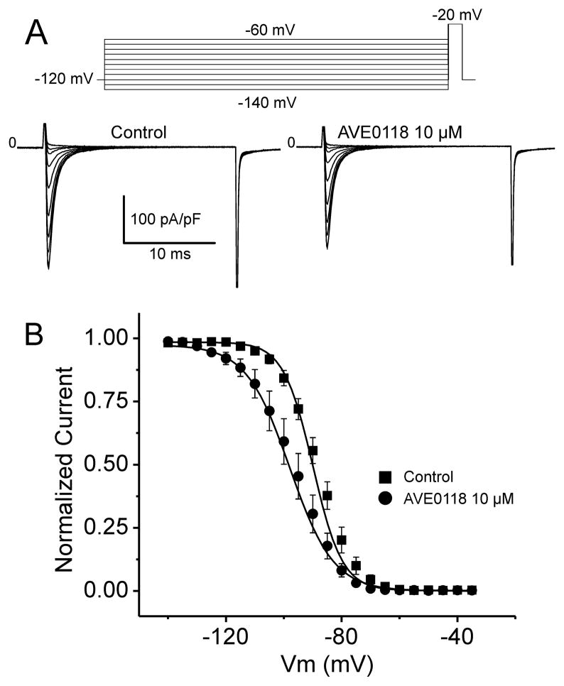
Figure 6. Effect of AVE0118 on Steady-state Inactivation of Cardiac Sodium Channels in HEK293 cells.
A: Representative Nav1.5 current traces recorded before and after 10 μM AVE0118. Currents were recorded using the protocol pictured in the inset. B: Peak Nav1.5 current was normalized to the maximum current recorded under control conditions or following 10 μM AVE0118 (-89.0 ± 0.55 mV vs. -95.96 ± 0.55 mV, respectively). Steady-state inactivation is plotted as a function of conditioning potential and fitted to a Boltzmann distribution. AVE0118 induces a significant shift in mid-inactivation voltage (V1/2; p<0.01). Mean ± SEM (n=7).

