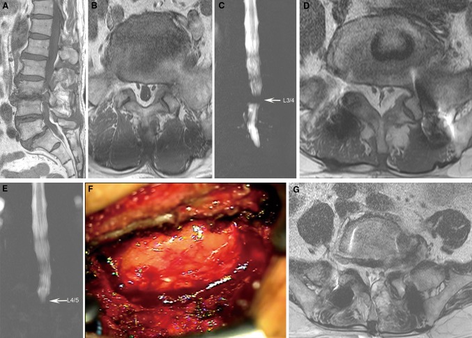
Fig. 1.
a T1-weighted sagittal MR image showing an extruded disc at L3/4 and isthmic spondylolisthesis at L5/S1. b T1-weighted axial MR image showing minimal epidural lipomatosis at L5/S1. c MR myelogram showing a complete block at L3/4. d Five months after checking initial MR image, T1-weighted axial image showing extensive epidural fat encasing the thecal sac circumferentially with a Y-shaped configuration at L5/S1. e MR myelogram showing a complete block at L4/5. f Intraoperative photograph showing abundant fat tissue encasing the thecal sac. g Postoperative T2-weighted axial MR image showing well decompressed the thecal sac and the nerve root with minimal epidural hematoma

