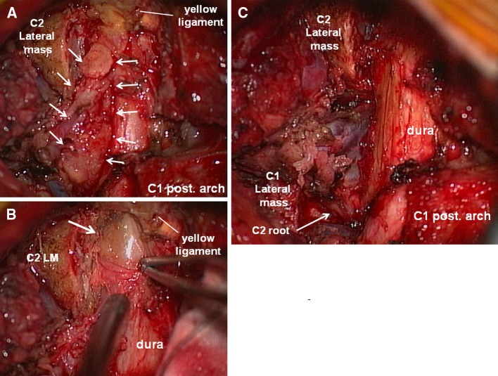
Fig. 2.
Intraoperative photographs of the patient. a Microscopic view after C1 and C2 hemilaminectomy on the right side. A waxy mass can be observed in the extradural space that extends laterally through the C1–2 intervertebral space (arrow). b A photograph after partial resection of the yellow ligament at the C2–3 level. A yellowish mass within the reactive outer membrane can be observed in the subligamentous extradural space without attachment to the yellow ligament. c Final microscopic view after resection of the mass lesion. The dural sac and right C2 nerve root (arrow) are decompressed and the ventral venous plexus can be observed

