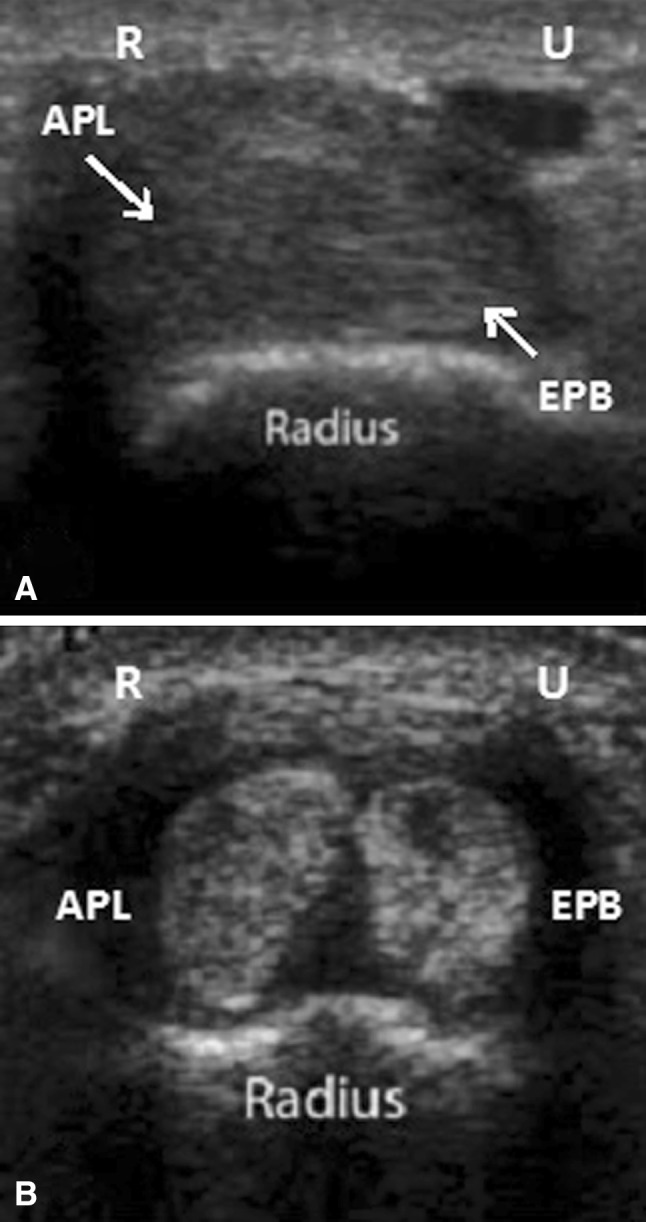
Fig. 2A–B.

Transverse axis sonogram of the first dorsal compartment show (A) a single compartment versus (B) two subcompartments. APL = abductor pollicis longus; EPB = extensor pollicis brevis; R = radial; U = ulnar. The presence of hypoechoic or anechoic material between the two tendons as seen in B also can be seen with a tendon sheath effusion and is not necessarily diagnostic of a septum. However, on injection there was no communication seen between two tendons.
