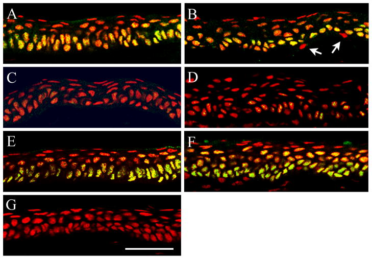
Figure 2.
Co-localization of ΔNp63 (green) and PI (red) to label epithelial nuclei (red) in the rabbit corneal epithelium following overnight contact lens wear. (A–B) In the non-lens wearing eye, ΔNp63 localized to the nuclei of all epithelial basal cells throughout the central corneal (A) and limbal (B) epithelium. Weaker expression was seen in epithelial nuclei in the wing cell layer. Expression was absent in all superficial cells. Occasional ΔNp63 negative cells were noted in the basal layer of the limbus (arrows). (C–D) 24 hours of PMMA lens wear decreased nuclear expression of ΔNp63 throughout the central corneal (C) and limbal (D) epithelium. (E–F) ΔNp63 expression and localization was unaltered after 24 hours of hyper-oxygen transmissible RGP lens wear compared to the control eye; central corneal (E) and limbal (F) epithelium. (G) Negative control, primary antibody omitted. Scale: 48 μm. Images representative of repeated experiments.

