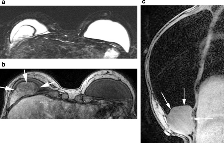
Fig. 12.
A 46-year-old woman with implant reconstruction. Axial a unenhanced T2W fat-saturated, b enhanced T1W, and c sagittal T1 silicone suppressed images show a circumscribed mass adjacent to the silicone implant in the inferior breast. The mass is bright on bright on T2W images and shows postcontrast enhancement. Biopsy proved this to be a spindle cell tumor

