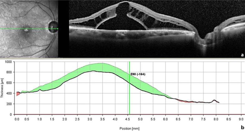
Fig. 2.
An SD-OCT scan through the fovea taken 4 weeks after the first surgical procedure (pars plana vitrectomy and ILM peeling) (a) shows a slight reduction of the inner retinal cysts near the disc, whereas the outer layer schisis-like separation and the retinal detachment did not improve. Moreover, an outer layer macular hole developed beneath the schisis. The decrease in central retinal thickness of 164 μm (b) is rather due to a displacement of the fluid than to reabsorption of it.

